Meet the Machines
Rare and astonishing equipment fuels science and medicine at UVA
Some are tiny. Others are massive. They’re helping doctors, students and researchers expand the limits of their fields. They’re UVA’s machines of science. Here’s a look at just a few.
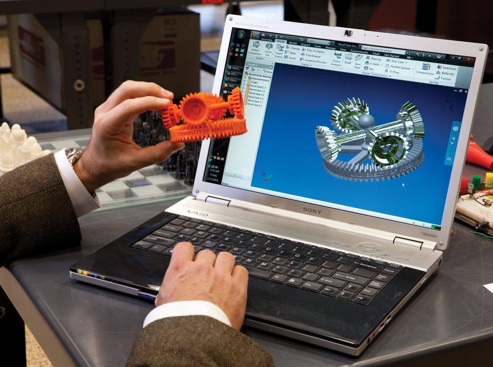
The 3-D Printer
Say you’re throwing a dinner party and find that you’re missing a salt shaker. What if you could go to your computer, download the design and “print” (create) one right then and there? Associate professor of engineering Gavin Garner (Engr class of ’06, class of ’09) says it won’t be too long before this becomes a reality.
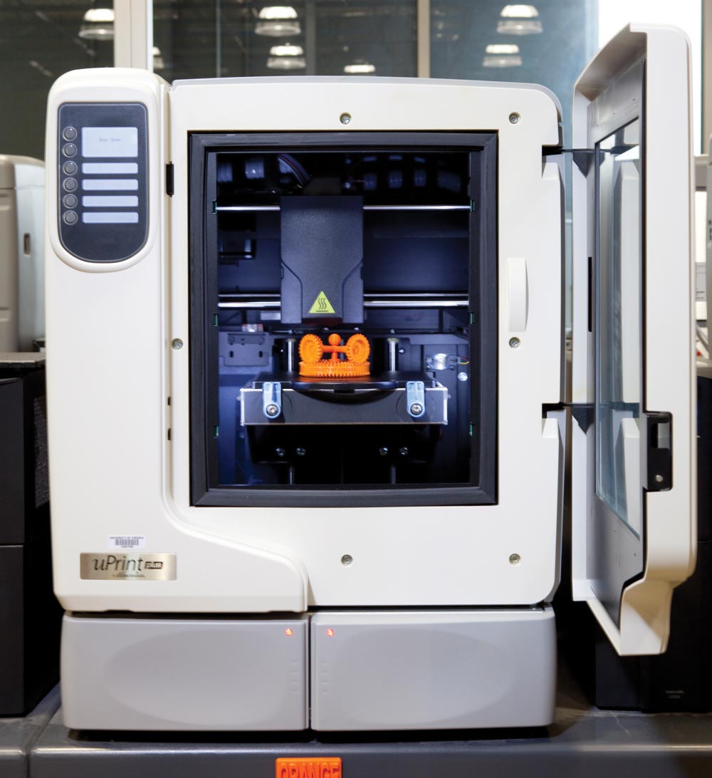
The recently opened Rapid Prototyping Laboratory in the Mechanical and Aerospace Engineering building houses six 3-D printers, or fused deposition modeling machines, each about the size of an oven, and one larger one, about the size of a small storage shed. “Whatever you can draw in three dimensions these computers can print,” says Garner.
The objects are made with ABS plastic, a material that melts at relatively low heat, around 300 degrees Celsius, and doesn’t create toxic gases.
The support material is a polylactic acid plastic. It’s used to fill in the gaps of an object being built. If you’re building an arch, for instance, the support material inside the opening allows the top to be finished.
The printer uses spools of plastic and a support material to make models. The plastic is laid down in fine layers with a number of quickly moving nozzles, while the support material is built around it, effectively allowing an object to be created midair. When the process is finished, the object is dipped in a chemical bath that strips away the support material, leaving the finished item.
“What we’re really doing with these machines is opening the doors to give our students and faculty the tools to be able to bring their dreams into reality,” says Garner.
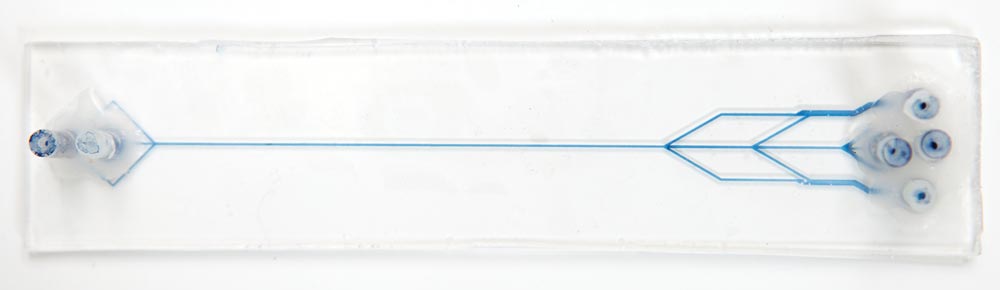
The Lab on a Chip
It usually takes up to two weeks for scientists to decode a genetic fingerprint. The “lab on a chip” provides the same results within an hour. A typical procedure involves an extraction, for instance getting your finger pricked. Analyzing that blood sample involves multiple steps in multiple places.
Graduate student Yiwen Ouyang uses transparent polyester sheets rather than glass to create inexpensive, disposable chips.
For a liquid sample to move through a chip, it must be attached to an ourside instrument (to the right of the frame), usually outfitted with a microscope, that controls what goes where.
“Lab on a chip stitches all of those together in a very small space,” says chemistry professor Jim Landers. “You can automate those processes, linked together and in miniature, and you can do it very, very fast.”
Under Landers’ guidance, students are creating labs on chips that they hope will one day be used in genetic and forensic testing as well as cancer prognoses.
Graduate student Dan Nelson extracts tumor cells from blood through micro channels on a plexiglass chip about an inch and a half wide and three inches long. The chip is etched with these channels, and then, using acoustic waves, the blood sample is pumped through them, traveling through various micro pipes and reservoirs where it can be analyzed and manipulated for results.
“Let’s imagine that Mrs. Jones goes in to see her doctor, and her lump is getting larger and larger,” says Landers. “The doctor sends her down to phlebotomy, where they’ll grab a couple thousand cells.” It will be days if not weeks, then, before the results come in. “Fast forward: If you have this chip in the office with the instrument, the doctor takes the cells himself, pops it in and tells Mrs. Jones to go get a cup of coffee. You’re on it today, not two weeks from now.”
The Gantry
Imagine you work for a car company and you’d like to test out a new color mix on an automobile, study how it appears in different kinds of light, without having to actually paint it and watch it drive around.
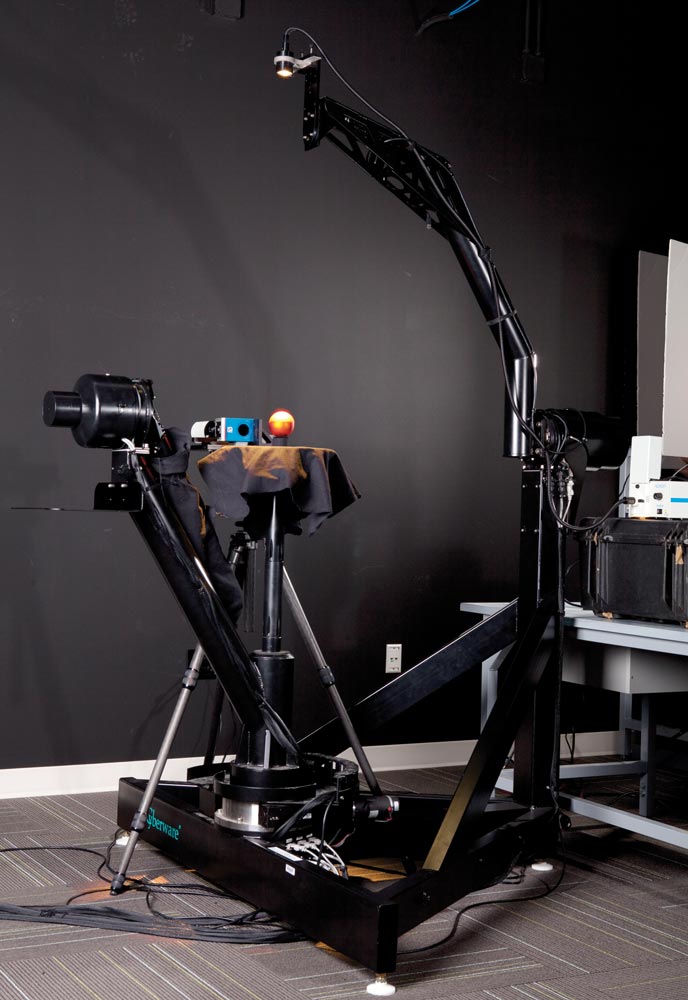
This is where the Gantry, a “robot for illuminating objects in a controlled way,” might come in, according to Jason Lawrence, an associate professor in computer science. One of four devices in the country, and originally developed at Stanford, the Gantry consists of two mechanical arms that swivel around a central platform.
An object painted with the new car color might be placed there, for instance, while the arms are fixed with different kinds of light. As they move around the platform, they simulate what the paint would look like on the car, providing “high-quality measurements of the appearance of an object,” says Lawrence, “and the way it scatters light.”
The Gantry is also used to help develop more effective 3-D scanners. “There are technical limits to what 3-D scanners can do, and this type of device helps researchers devise new ideas of how to improve them,” says Lawrence. It can support research in industrial design, architecture, manufacturing and digital humanities, any area that could be served by “predictive rendering,” or a simulation of how something is going to look.
The Tomo Linear Accelerator
Below UVA’s Emily Couric Clinical Cancer Center, in thick-walled underground rooms built with lead bricks and sealed with heavy doors to shield radiation, is one of the world’s most advanced cancer-fighting machines. The TomoTherapy Linear Accelerator can deliver doses of radiation to a tumor with such pinpoint accuracy that surrounding healthy tissue is unaffected.
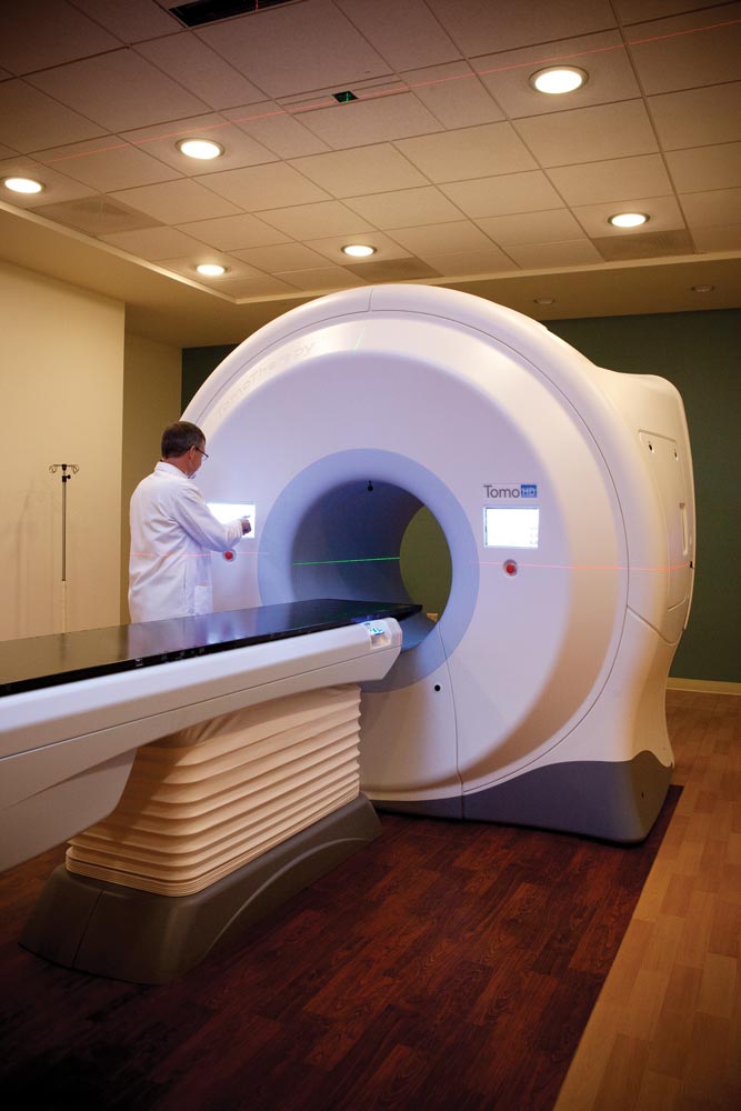
The patient slides into the machine and a CT scan is taken with a helical rotation x-ray beam that swivels 360 degrees around the patient. This scan is compared with previous scans to provide a 3-D image, highlighting any changes in the tumor that have taken place. The patient is positioned accordingly, and treatment can begin. “Twenty-five years ago you got two fields,” says Dr. Jim Larner (Med class of ’80), professor in the department of radiation oncology, “one from the front, one from the back.”
One of the feats of engineering in this machine are the 64 tungsten leaves that block or unblock the radiation during treatment, depending on the size and shape of the tumor. The same helical rotation beam is used to create tiny beam elements, known as beamlets, that can better target the tumor. If a person is being treated for a tumor in his prostate, for example, the technology keeps the dose from affecting the bladder or rectum, usually a complication with this kind of treatment.